Health Innovations
Innovative Imaging Blends Light and Sound to Track Real-Time Brain Oxygen Levels
A dual imaging technique developed at Washington University tracks brain oxygenation in real time, offering new insights into vascular conditions that disproportionately affect Black communities.
Published September 22, 2026 · HealthHBCU Newsroom
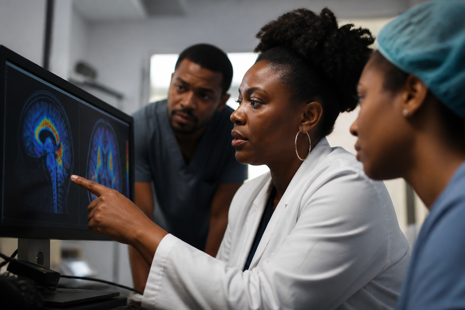
A new imaging technology developed at Washington University in St. Louis offers an unprecedented look inside the living brain, allowing scientists to monitor blood flow and oxygen delivery in real time. The approach, which merges optical light and acoustic ultrasound waves, could help biomedical researchers better understand how brain cells and blood vessels interact—and what goes wrong when that partnership fails in conditions like stroke, vascular dementia, and cognitive decline.
For Black communities, where vascular health challenges and stroke rates are disproportionately high, advancing our understanding of cerebral blood flow is critical. Black Americans are up to twice as likely to have a stroke as white Americans and face higher mortality rates from cardiovascular and cerebrovascular diseases. Emerging technologies that illuminate the precise mechanics of brain oxygenation provide essential tools for the scientists and clinicians working to eliminate these deep-seated health disparities.
Combining Light and Sound to See the Brain
Developed by Song Hu, an associate professor of biomedical engineering at Washington University, the novel system combines optical resolution photoacoustic microscopy with high-frequency ultrasound imaging. While traditional brain imaging methods often require a trade-off between spatial resolution and depth, this dual-modal technique captures high-contrast images of microvascular networks alongside structural maps of surrounding tissues.
By tracking how hemoglobin absorbs light at different wavelengths, the system calculates oxygen saturation levels in individual microscopic blood vessels without invasive dyes or contrast agents. This real-time capability allows researchers to observe dynamic changes in oxygen delivery as neural tissue demands more energy during brain activity.
Addressing Vascular Risk and Brain Health Disparities
The coupling between neural activity and local blood flow, known as neurovascular coupling, is fundamental to maintaining healthy brain function. When chronic high blood pressure, diabetes, or systemic inflammation damages fragile capillaries, this critical mechanism degrades, leaving brain tissue vulnerable to hypoxia and cellular death.
Because hypertension and metabolic disorders disproportionately affect Black adults, understanding microvascular decay is a major priority for health equity researchers. By providing a clearer picture of how vascular risk factors compromise brain oxygenation over time, tools like Hu's imaging platform can help translational scientists design targeted therapies and early interventions to preserve cognitive function in high-risk populations.
The Role of HBCU Researchers in Bioimaging Innovation
Translating novel biomedical tools into clinically effective solutions requires a diverse workforce of engineers, neuroscientists, and physicians. Historically Black Colleges and Universities with strong engineering and pre-medical programs—such as Howard University, North Carolina A&T State University, and Morgan State University—are playing a crucial role in cultivating the next generation of Black leaders in bioinstrumentation and neurobiology.
Ensuring that Black researchers are at the forefront of developing and applying advanced imaging technologies is essential for shaping research agendas that prioritize health equity. As tools for observing neurovascular health become more sophisticated, early-career scientists trained at HBCUs will be instrumental in deploying these innovations to address the specific health burdens impacting underserved communities.